X-ray manual : U.S. Army . <*5 lit w Fig. 3. Position for first exposure.. Fig. 4. Position for second exposure.200 HEAD EXAMINATIONS 201 moved from the front plane of the
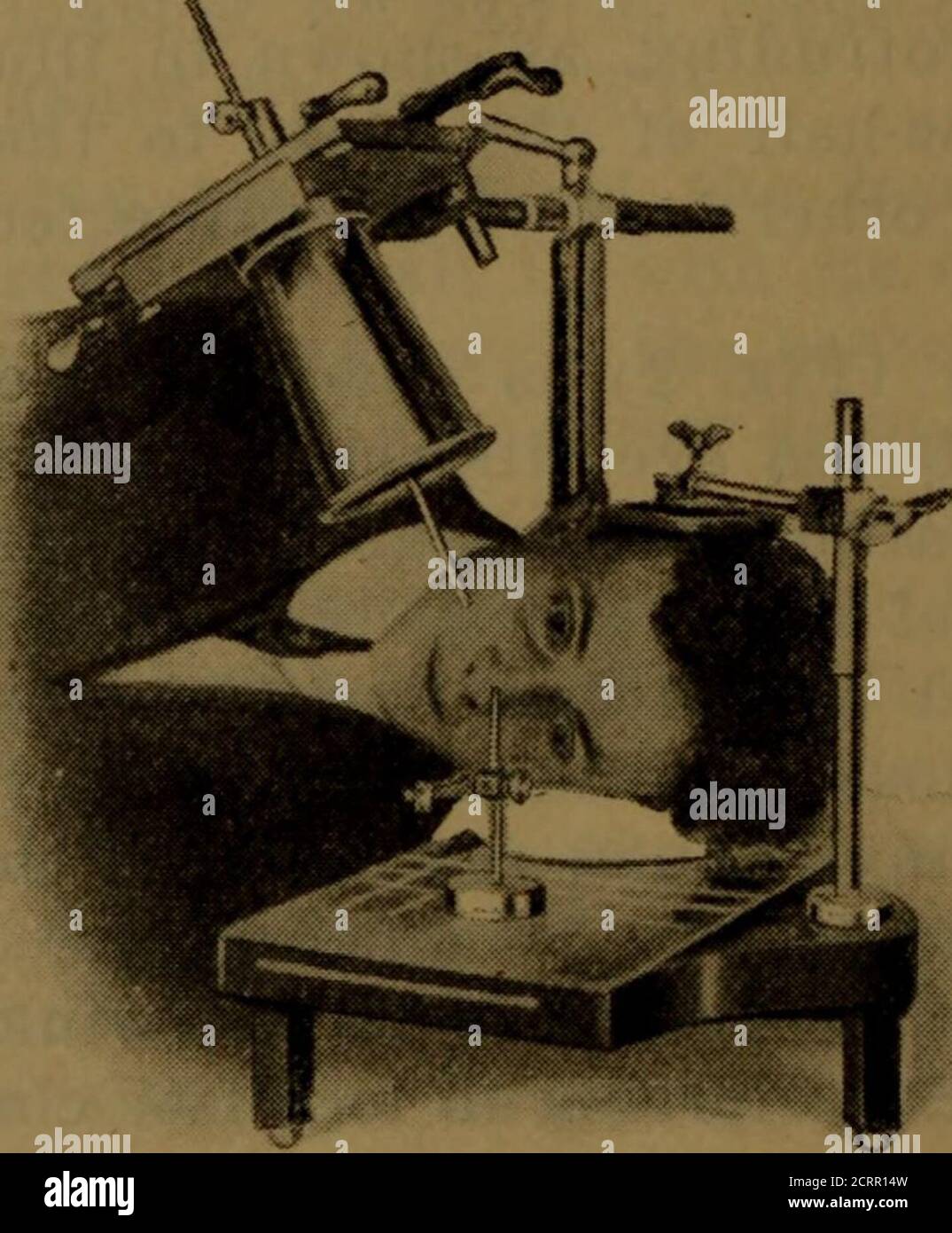
Download this stock image: . X-ray manual : U.S. Army . <*5 lit w Fig. 3. Position for first exposure.. Fig. 4. Position for second exposure.200 HEAD EXAMINATIONS 201 moved from the front plane of the cornea, and it shouldalso be borne in mind that the front of the cornea is 10millimeters in front of the shadow of the indicator-ball, asshown in your negatives. The tube is now centered overthe localizing ball and cone so that the shadows of thetwo will coincide (Fig. 3). Some object, such as a candle or a piece of whitepaper, that can be readily seen by the patient, should beplaced in alignment with the sights of the - 2CRR14W from Alamy's library of millions of high resolution stock photos, illustrations and vectors.

CDA Journal - March 2021: Oral and Maxillofacial Reconstruction by California Dental Association - Issuu

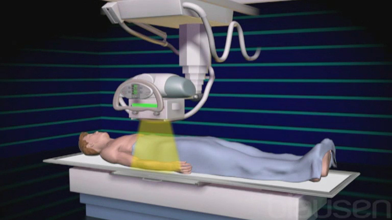
Radiographic Exposure Technique

Concussion Management Policies and Procedures by Andrew Stabell - Issuu
X-Rays - Special Subjects - MSD Manual Consumer Version

United States Army X-Ray Manual: United States. Surgeon-General's Office: 9781141999200: : Books
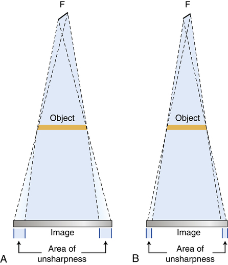
Radiographic Exposure Technique

Travma 2012-3 by KAREPUBLISHING - Issuu

AER 9.3 by Radcliffe Cardiology - Issuu

Operational guidelines and procedures for measuring the real size of the world economy part 1 by World Bank Publications - Issuu

First aid emergency medicine by Rosana Rosa - Issuu

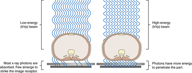
Radiographic Exposure Technique
Radiographic Exposure Technique
X-ray manual : U.S. Army . <*5 lit w Fig. 3. Position for first exposure.. Fig. 4. Position for second exposure.200 HEAD EXAMINATIONS 201 moved from the front plane of the

lekarz wojskowy 2016 02 book kor en by Medycyna Praktyczna - Issuu

WHO Laboratory Manual for the Examination of Human Semen by Shah Tarang - Issuu