shows SEM micrographs of the surfaces of the silica gel as a function
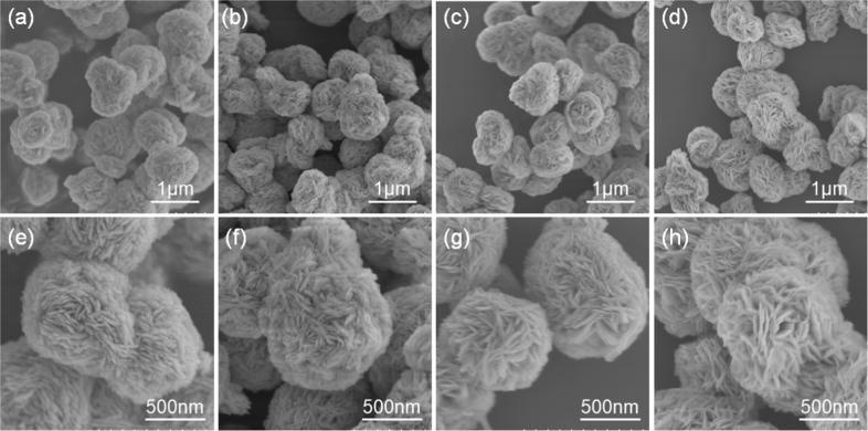
Download scientific diagram | shows SEM micrographs of the surfaces of the silica gel as a function of the soaking time in SBF. It can be seen from that apatite started to precipitate on the silica gel, taking the form of tiny spherulites about 5 days after the gel was soaked in SBF. The spherulites increased in both number and size with increasing soaking time, and completely covered the surface of the silica gel within 14 days. Fig. 4 shows higher-magnification SEM micrographs of the apatite formed on the silica gel soaked in SBF for 14 days. It can be seen from Fig. 4 that each spherulite consists of a large number of flakes. Some spherulites were formed not only directly on the surface of the silica gel, but also on the surfaces of other growing spherulites (Fig. 4a) or at their interfaces (Fig. 4b). This indicates that the front of a growing apatite from publication: Process of Formation of Bone-Like Apatite Layer on Silica-Gel | It has been proposed that a hydrated silica plays an important role in forming a biologically active apatite layer on the surfaces of bioactive glasses and glass-ceramics in the body. Recent experiments have shown that a silica hydrogel actually induces apatite formation on | Apatites, Silica Gel and Bioactive Glasses | ResearchGate, the professional network for scientists.

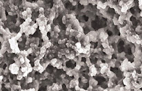
SEM image of silica gel obtained at different magnifications

The application of silica gel synthesized from chemical bottle

SEM images for: (a) Silica gel RD-2060, (b) HKUST-1, (c) Fe-BTC

Atomic force microscopy - Wikipedia
Superhydrophobic surfaces review: Functional application
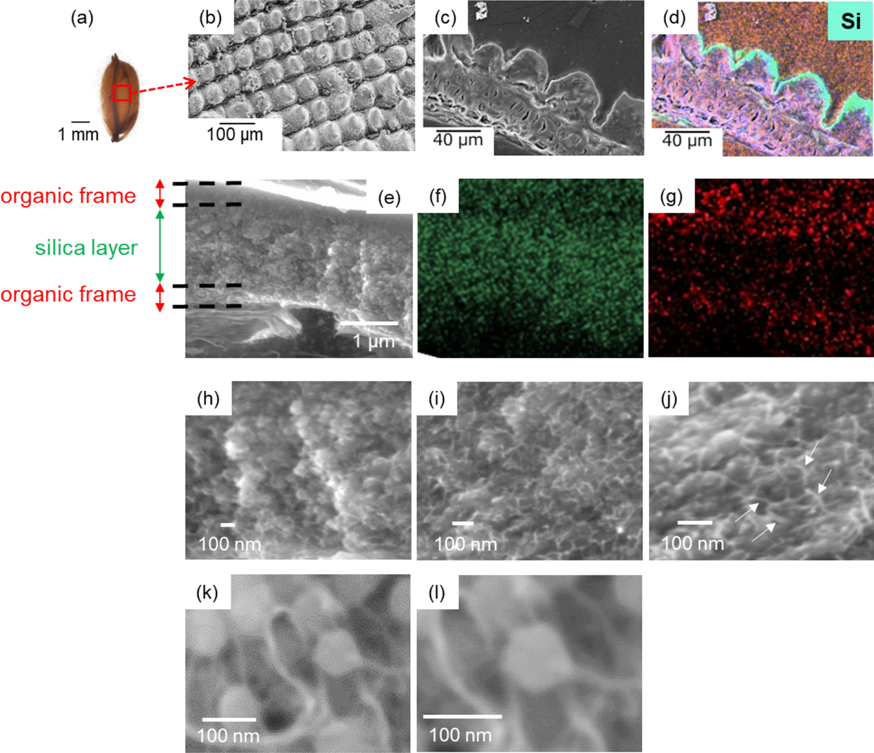
Cellulose intrafibrillar mineralization of biological silica in a

Gels, Free Full-Text
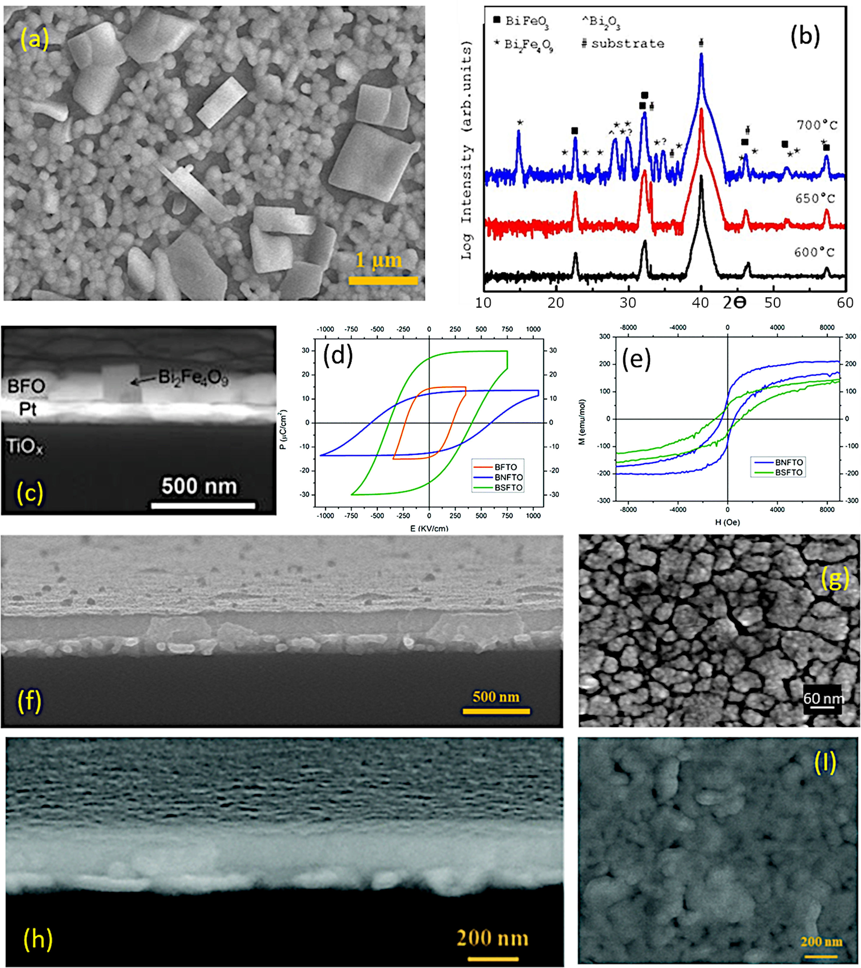
Thin film processing of multiferroic BiFeO3: From sophistication
Micrograph of the Month Journal of Sol-Gel Science and Technology

Silica gel with continuous macropores prepared from water glass in
Silica-gel Manufacturer